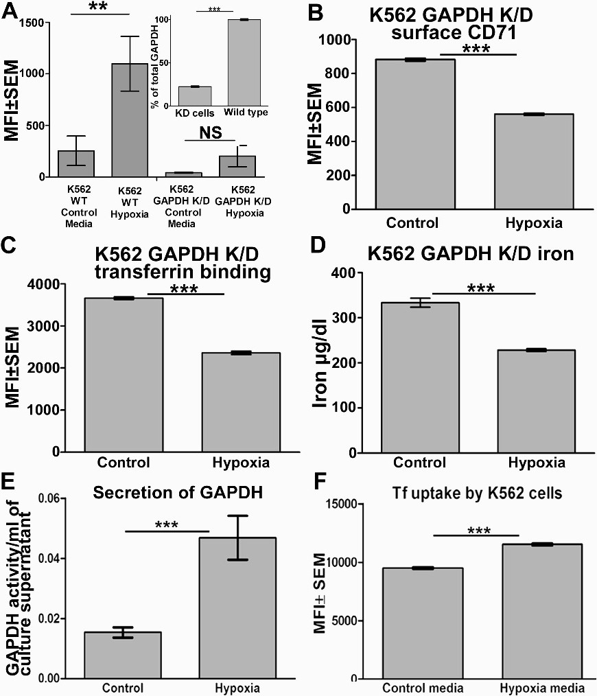
Fig. 4. Knockdown of GAPDH confirms its role in cellular acquisition of Tf and Tf bound iron during hypoxia. (A) Unlike wild type cells the GAPDH K/D cells do not show any increase in their surface GAPDH recruitment upon hypoxic exposure. K562 wild type and GAPDH knockdown cells were stained for surface GAPDH after exposure to hypoxia for 24 hrs and analyzed by flow cytometry, control cells were maintained in regular normoxic culture conditions, ** p<0.001, NS p>0.05, n=104. (A inset) Total cell GAPDH is significantly decreased in knock down cells as compared to wild type cells. p<0.0001, n=3. (B) As in case of wild type cells, hypoxia causes a significant decrease in CD71 expression on the surface of GAPDH K/D cells: K562 GAPDH knockdown cells were stained for surface CD71 (TfR1) after exposure to hypoxia for 24 hrs and analyzed by flow cytometry, control cells were maintained in regular normoxic culture conditions, p<0.0001, n=104. (C) Hypoxia treated GAPDH K/D cells are incapable of increasing their transferrin capture: K562 GAPDH K/D cells exposed to hypoxia for 24 hrs or maintained in normoxic culture conditions (control) and then assayed for transferrin binding by flow cytometry. A significant decrease in transferrin capture is evident, p<0.0001, n=104. (D) GAPDH knockdown cells are unable to maintain their iron uptake upon hypoxic exposure, p<0.05, n=3. (E) Hypoxia causes enhanced secretion of GAPDH. Increased levels of secreted GAPDH are present in culture supernatant of hypoxic K562 cells, p<0.0001, n=4. (F) Culture supernatant of hypoxic cells enriched in secreted GAPDH effects more transferrin uptake into cells. Fresh cells maintained in culture were incubated with 10 µg of TF-A647Tf re-suspended in 200 µl media supernatant from hypoxia exposed or control cells and evaluated for internalization of transferrin by flowcytometry, p<0.0001, n=104.